Software for High Resolution Electron Microscopy
| home |
| updates |
| demos |
| structures |
| contact |
MacTempasX
MacTempasX Version 2 represent a major upgrade of
MacTempas. The program has been expanded to include a
huge number of new features. In addition to the basic
image/diffraction HRTEM capabilities, STEM and CBED calculations
have been included. The program now includes both basic
and advanced image processing capabilities as well as
quantitative image comparison and structure refinement.
Short Highlight of MacTempasX Features |
|
| Example screen shots from MacTempasX |
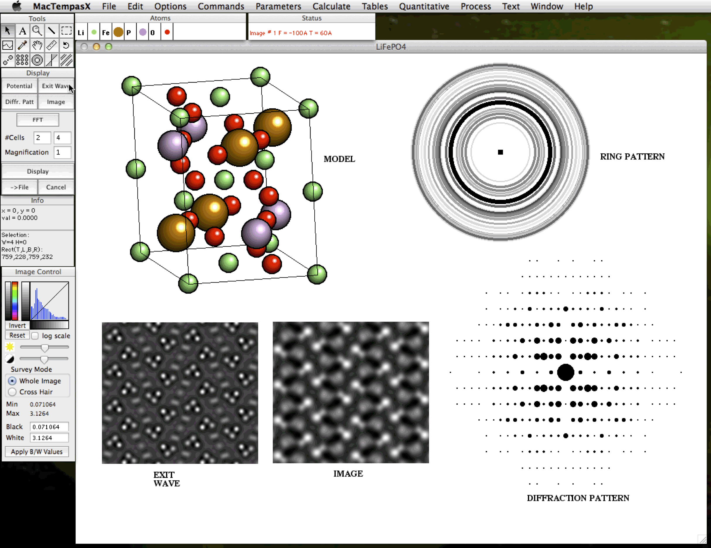
Main Display Window showing some examples of calculated data from a given atomistic model
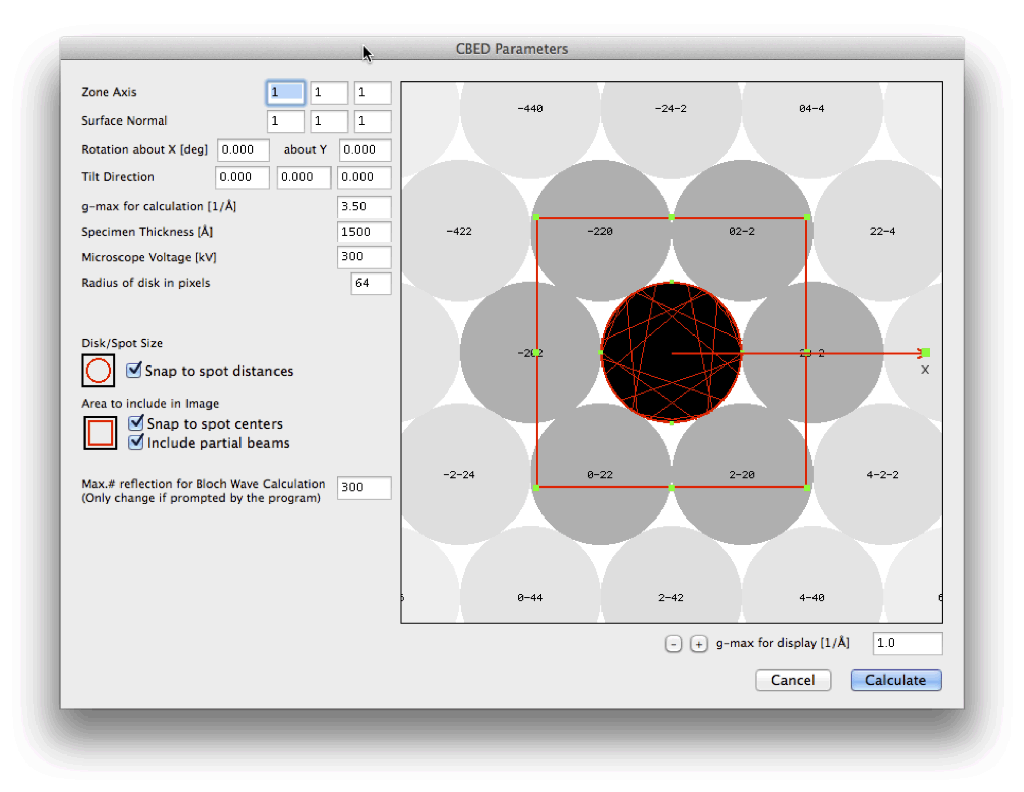
Starting point for computing a CBED Pattern
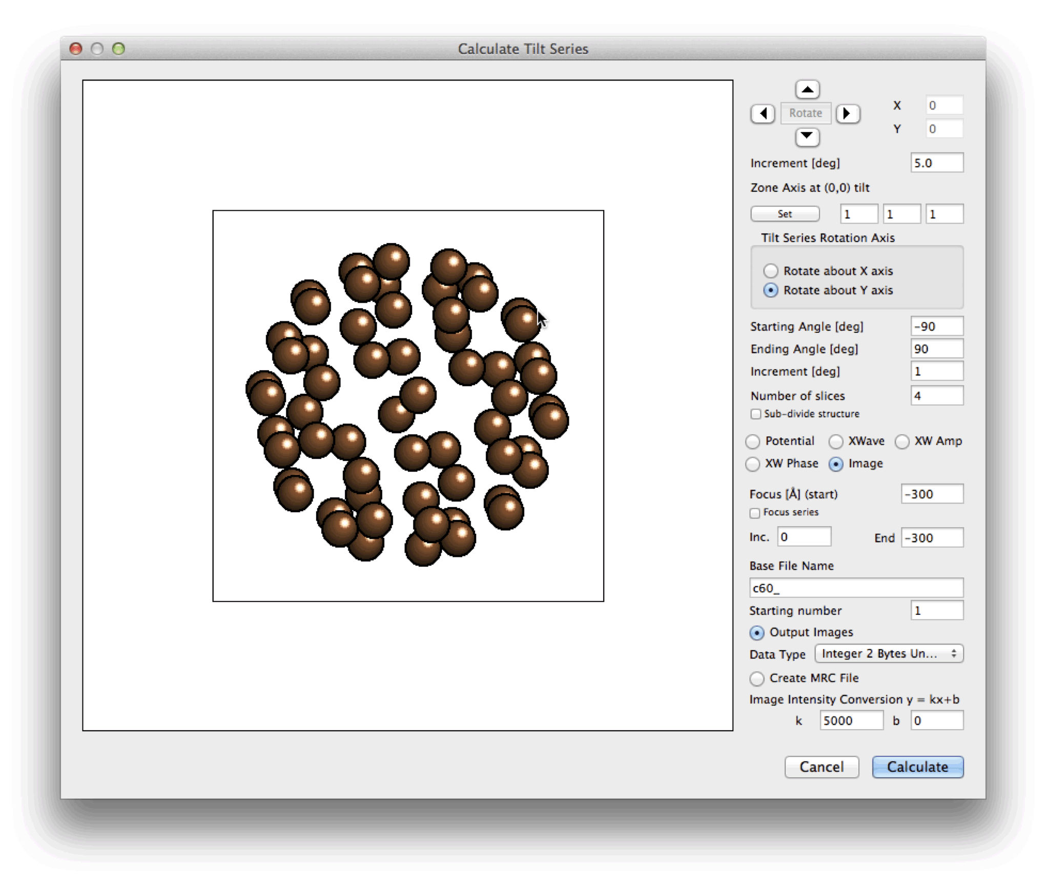
Starting point for computing a Tomographic Tilt Series
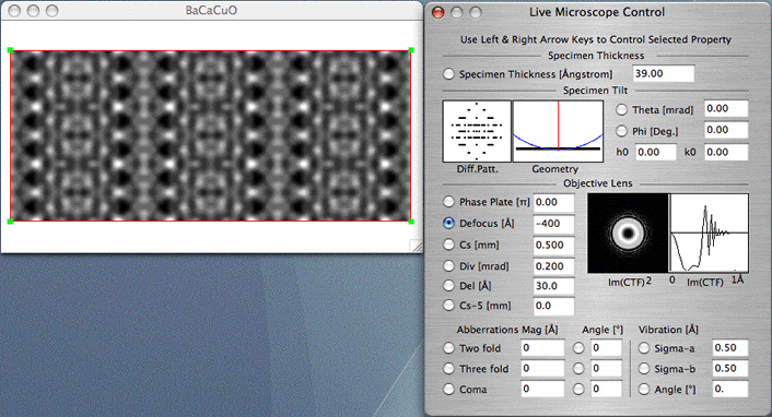
A Simulated Image with a "Live Microscope Control" The Image is updated "Live" as new input parameters are given
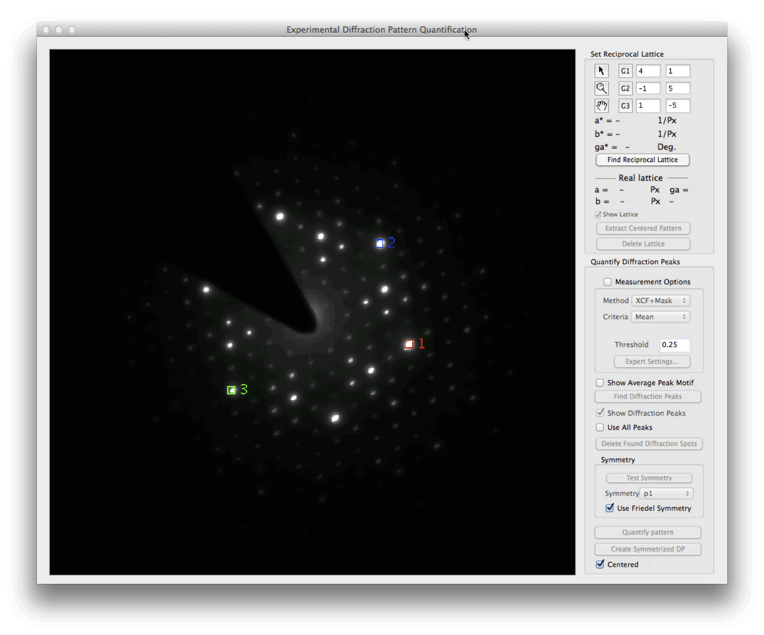
The Control Window serving as a starting point for extracting quantitative data from experimental diffraction patterns
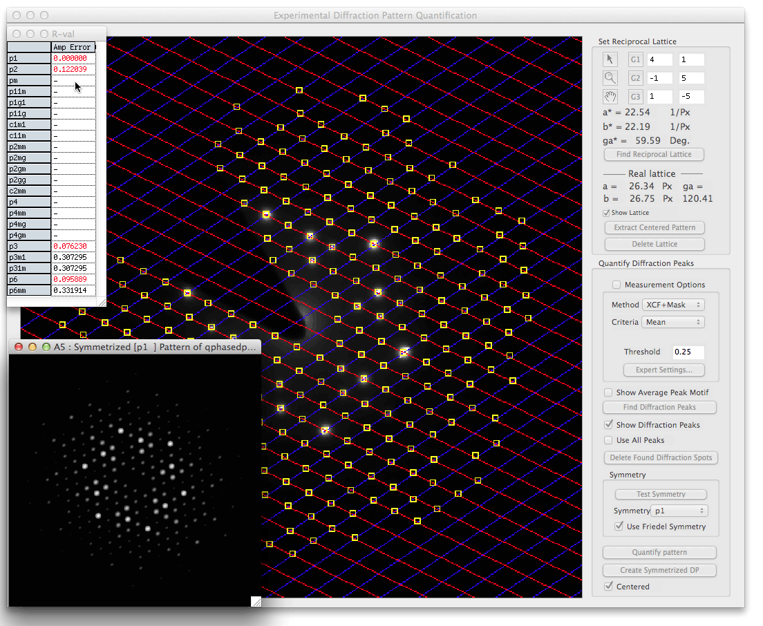
Peak information extracted from the diffraction spots at the determined lattice
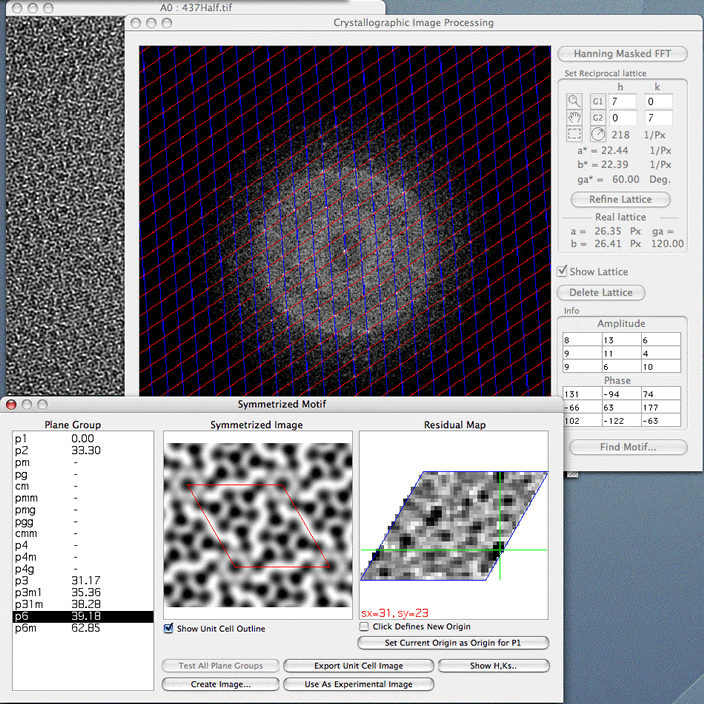
Crystallographic Image Processing using symmetrized data from the 2D-Periodic Unit Cell
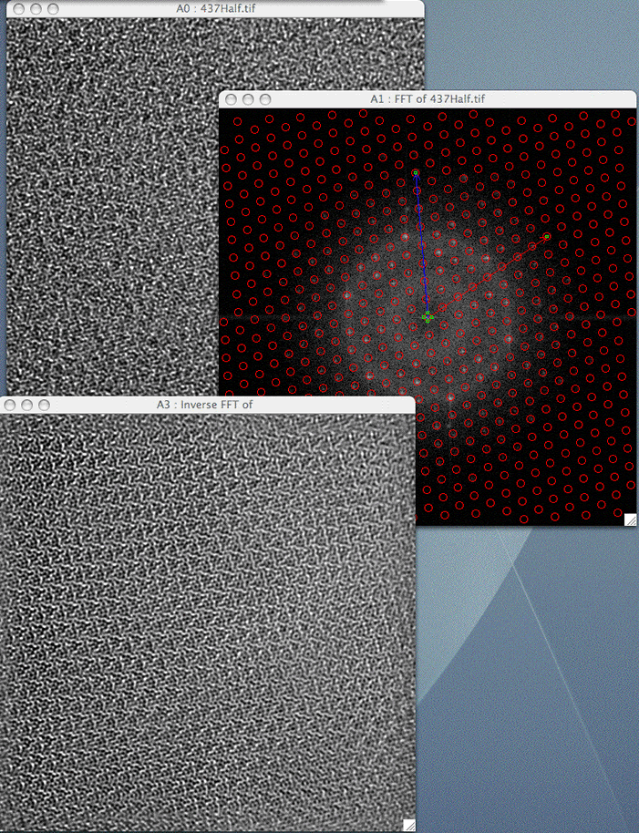
Fourier Image Processing using a periodic lattice mask
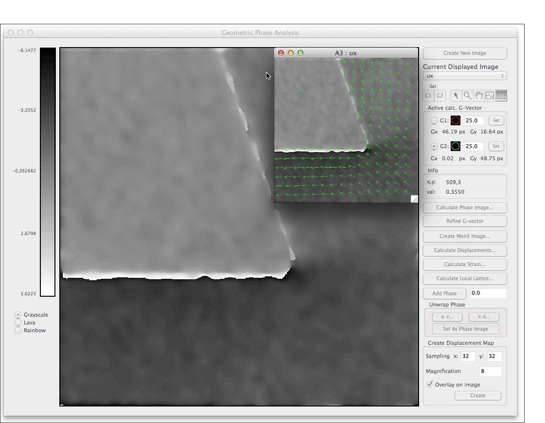
Geometric Phase Analysis using data from information around diffracted spots to determine lattice deviations and strain
Geometric Phase Analysis showing a gray level depiction of the deviations from a reference lattice in the x-direction together with a vector map showing the 2D deviations from the same lattice.
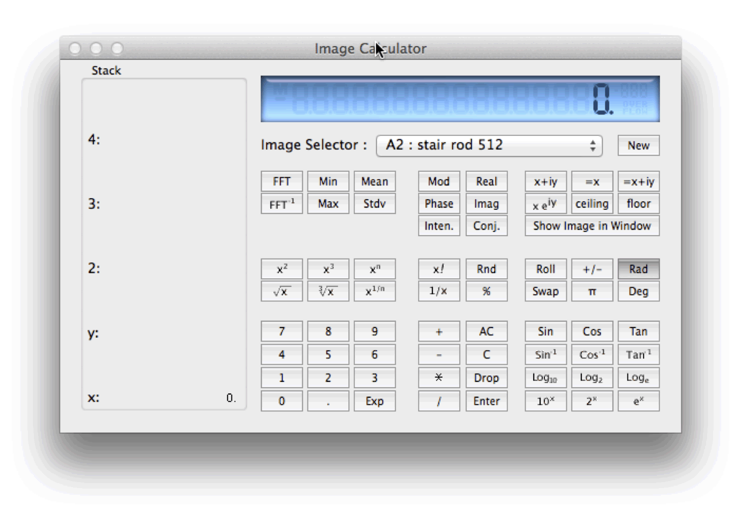
The built in Reverse Polish Notation Calculator which operates on real and complex images as well as real and complex numbers
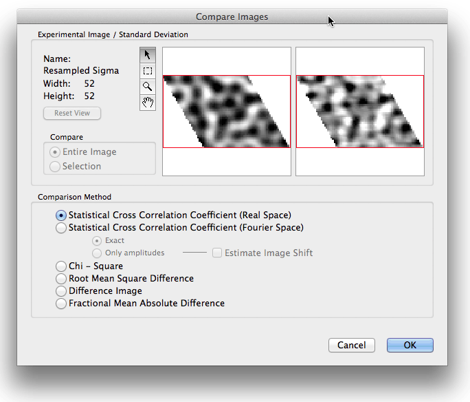
Starting point for quantitative matching between experimental data with simulation using a number of criteria for the quantitative fit. The quantitative fit can also be used to refine a structure by minimizing the discrepancy between calculation and experiment
|
|
|